
You can see Chlorella and Spirulina with a basic school microscope. At 400x magnification, Chlorella shows as clear green spheres about 2 to 10 micrometres across, and Spirulina as blue-green coils with a visible cross-banding along the filament. A few minutes under the lens tells you more about your culture’s health than days of staring at the jar.
What you need
- A compound microscope with 10x eyepiece and 10x, 40x objectives (100x and 400x overall).
- Glass slides and cover slips.
- A dropper or pipette.
- Light: the built-in lamp is fine.
- Optional: a smartphone adapter for photos, and a counting chamber if you want cell numbers.
Preparing a sample
- Stir the culture and take a drop from the middle, not the top or bottom.
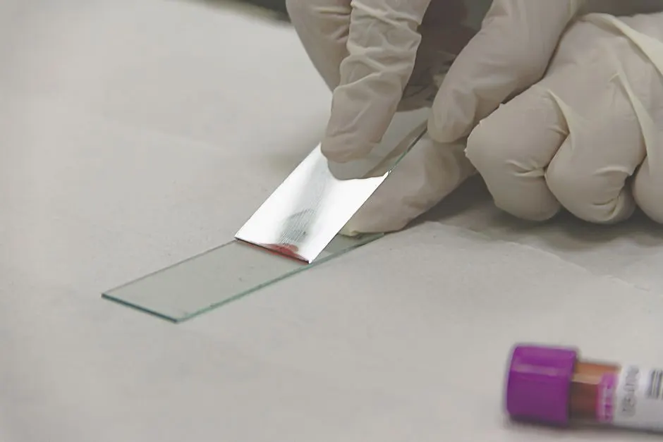
- Place one drop on a slide. If the culture is very dense, dilute it with a drop of clean water.
- Lower a cover slip at an angle to avoid trapping bubbles.
- Start at 100x to find the focus, then move to 400x.
What you will see
Chlorella
At 400x, cells are small, round and uniformly green, often with a paler centre. Some show two or four small cells packed inside a wall, which are autospores about to be released. Empty, pale rings are old walls. See how Chlorella reproduces for what you are seeing.
Spirulina
At 100x, filaments appear as green-blue spirals, often a few turns long. At 400x, you can see the cells as short segments stacked along the filament. Healthy filaments are swimming slowly with a gliding motion. Details of the structure are in how Spirulina grows.
Healthy or contaminated?
| You see | Likely meaning |
|---|---|
| Even green cells, little else | Healthy culture |
| Many small dots and rods that move in jerks | Bacteria; check smell and overfeeding |
| Larger, oval cells swimming fast | Ciliates or other protozoa grazing |
| Cells with grey or empty interiors | Dying or dead cells |
| Dark clumps with strands | Fungal growth or debris |
If you find grazers or heavy bacteria, restart from a clean reserve and review your handling; preventing contamination lists the usual causes.
Counting cells
A counting chamber, also called a hemocytometer, lets you count the cells in a known volume. That gives you a number you can use to compare batches or to dose a feed precisely. It is optional for hobby use, but helpful in small-scale production.
Getting the best image
- Lower the condenser or close the iris slightly. Transparent cells show better with a little contrast; fully open light washes them out.
- Use a thin sample. One drop under a cover slip, with the excess wicked away with tissue, gives a single layer of cells.
- Focus with the coarse knob at 100x first. Then switch objectives and only use the fine knob.
- Clean the lens. A smudge on the objective looks like a mysterious object in every field.
- Be patient with motion. Spirulina glides; wait a moment for filaments to settle in the field.
Counting with a grid
If you want numbers, a hemocytometer is a glass slide with a ruled grid etched in it, with known volumes between the lines. The procedure is simple:
- Dilute the culture so that cells are easy to count, for example 1 part culture to 9 parts water for dense Chlorella.
- Fill the chamber with the diluted sample and let cells settle for a minute.
- Count cells in several large squares at 400x, skipping those that touch two chosen edges so they are not counted twice.
- Calculate: average cells per square multiplied by 10,000 (for the standard 0.1 µL squares), multiplied by the dilution factor, gives cells per millilitre.
A dense Chlorella culture lies in the range of 10 to 50 million cells per millilitre. Counting a culture over several days gives you a growth curve, comparable to the one in the growth phases of an algae culture.
Other things you might see
| Object | What it looks like | Meaning |
|---|---|---|
| Diatoms | Golden-brown glassy boxes or boats | Wild contaminant; common in tap water |
| Rotifers | Larger animals with a wheel of hairs | Grazers; reduce culture |
| Ciliates | Oval cells, rapid swimming, fine hairs | Grazers; a sign to restart |
| Bacteria | Tiny rods and dots, shimmering | Normal in small numbers; bloom means trouble |
| Crystals | Sharp, angular shapes | Dried salts from the medium; harmless |
| Air bubbles | Dark rings with bright centres | Artefact of a poorly made slide |
Recording what you see
A phone held to the eyepiece takes usable photographs. Save one image per jar per week, and put the date and magnification in the file name. Combined with your notes in the culture log, they provide a visual history and are helpful when you ask others for help diagnosing a problem.
Common questions
Is a 40x microscope enough?
It is enough for Spirulina coils. For Chlorella, 400x is better.
Can I use my phone?
Yes, with a clip-on lens or by holding the phone to the eyepiece. Image quality is good enough to compare samples.
Do I need stains?
No. Both organisms are pigmented and visible without staining.
What magnification do I need to see bacteria?
Individual bacteria need 1000x with oil immersion. At 400x you see them only as a shimmer or as moving dots.
Can I use a USB microscope?
Yes, a good one can show Spirulina coils at the highest magnification, and it makes recording easy.
Ready to grow your own?
Live starter cultures, grown with care and shipped ready to thrive.
Spirulina PlatensisChlorella VulgarisVisit the shopGet new guides by email, and 10% off
Subscribe for 10% off your first order, plus our latest articles by email. No spam, unsubscribe any time.
Keep reading
What Is Phycocyanin? The Blue Pigment in Spirulina
Chlorophyll in Algae: How Microalgae Capture Light
Leave a Reply